


- This is a blood drop with a clotting disorder.

“e” upside down

Compound Microscope Mirror, HD Png Download - vhv

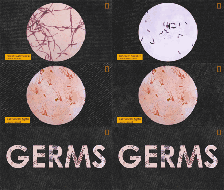
exprmnt 003 - @nugeish on Instagram


Gnetum leaf under microscope


- 10 Best Travel Destination In USA

Strawberry DNA

Premium Photo | Close up of Microscope in a laboratory


- [JLC] Getting 75-year-old Radium lume to glow again!

Biology cells microscope



- Bottle potion

Bacteria under microscope aesthetic colorful

Strawberry DNA


- All about Henna

I love 🔬

Onion cells


- Adhocism


Microscope virus close up. Created with Generative AI stock photo

- All about Henna


- Small Animal Sci

biology

biology


- Copper wire

Microscopic artifact


- Alzheimers or other Dementias:Minimize (or Prevent)

Gnetum leaf under microscope

Premium Vector | Microscope

- Aftermath of the tear gas canister incident.


Mikroskop | Premium wektor wygenerowany przez AI

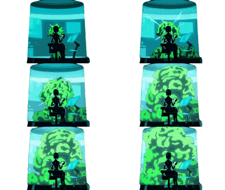
- A fiber from my lab coat fell in my sample this morning. It was tied in a tiny knot.


Download free image of Microscope microscope white background magnification. by Ing about texture, hand, aesthetic, light, and technology 12441784

- Origami lights


Ascaris cross section


- Forbidden corn nuts

Ascaris cross section

Medical Microscope PNG Transparent, Medical Microscope Hand Painted Medical Microscope Cartoon Medical Microscope Fine Medical Microscope, Medical Microscope Illustration, White Microscope, Medical Microscope PNG Image For Free Download

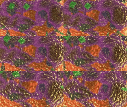
- Molecular Gastronomy

Onion cells

Red Microscope stock photo. Image of technology, education - 11443538


- A cell panda under an electron microscope


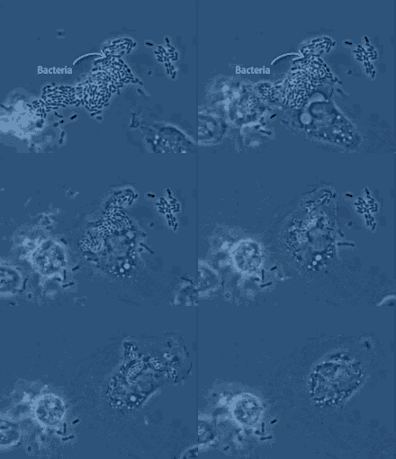
- Had to share didn’t realize chalk was so cool


- Free Material Pack: Stone And Bricks

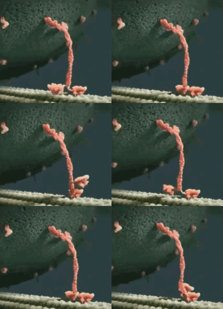
- Staying up late with friends trying to see a nipple through the scrambled channels.


- This electron microscope lets you literally see the atomic bonds like on a diagram

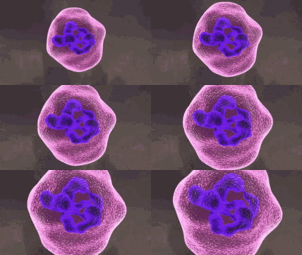
- Contemporany art


- Earthworm cross-sections from X-ray micro-CT scans [2244x3403]

- diy music


- This is the smallest 3D printed object and it is so small that it can fit on a needle

- [ art ] ceramic


- Pattern, Texture, Material

- 3d printed dress


- There’s a chance that when you’re pregnant, instead of growing a baby and a healthy placenta in your uterus, you grow a gestational aberration called a hydatiform mole. Here’s one where the mole has completely filled up the uterus and obliterated the cavity.


- Agates (1)

- Mitral Valve Prolapse

- Biology


- Illustration - Characters Design


- ...SHHHH...ITS A SECRET!


- 1900s

- The Near-Earth Object Camera (NEOCam) Infrared Sensor [2160 x 1439]


- Anatomy and Physiology


- Nikon small world


- These geologic formations on Venus that look like a monster head


- Anatomy and surgery


- Nano Particles at x100000 zoom

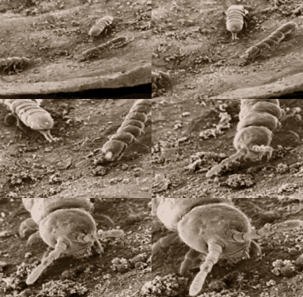
- BLACK & PURPLE!


- Mammatus clouds

- This ice cave in my local snow bank


- adrian

- Fatty Liver Symptoms


- Alicia Tormey


My favorite playground... Ready to Go! 👏🏻 #ICONProducts - @curlychiara on Instagram


- Amazing YOU!

- Plant Cell Photos


- Crop Circles

- anatomy sketch


- My aunts in-floor heating thermal image


- Chaotic terrain on Moon Europa, Jupiters Ocean world. © NASA/JPL/ASU [Xpost from r/cosmicporn]

- Brain & Nervous System


- Adam and Eve, Roman Opalka, Etching, 1968


- Cell & Microbiology


- Kids Party Supplies


- Plant Science


A carotid-cavernous sinus fistula (CCF) is an abnormal connection between an artery in your neck and the network of veins at the back of your eye. These veins at the back of your eye transport blood from your face and brain back to your heart and are located in small spaces behind your eyes called cavernous sinuses. Sometimes an abnormal channel forms between these veins and one of the internal or external carotid arteries that run up each side of your neck. This formation happens as a result of a small tear that sometimes occurs in one of the carotid arteries. If the tear occurs near the veins in the cavernous sinus, an abnormal channel may form between the artery and the network of veins, through which blood may flow. This is called a fistula. A fistula can raise the pressure in your cavernous sinuses, which may compress the cranial nerves located around the cavernous sinuses. This compression may damage the nerve function, which is to control your eye movements. These cranial nerves also allow you to experience sensation in parts of your face and head. The increased pressure caused by the fistula can also affect the veins that drain your eye. This can cause symptoms such as eye swelling and abnormal vision. Symptoms of Carotid-Cavernous Sinus Fistula Indirect CCFs tend to cause fewer, less serious symptoms. This is because of their relatively low rate of blood flow. Direct CCFs usually require more urgent attention. For both types, symptoms may include: •a bulging eye, which may pulsate •a red eye •an eye that protrudes forwards •double vision •loss of vision •an audible swish or buzz coming from your eye •weak or missing eye movements pains in your face •ringing in your ears •headaches •nosebleeds Case by @kmkoptometrypro - @optometry.case on Instagram


*EDITED* NEW BLOG POST ••• Wood is a wonderful material to be observed under the microscope. A tree trunk with its 3-dimensional structure needs 3 cutting planes to be fully understood: - Cross section - Image 1&4 - Radial cut - Image 3 - Tangential cut - Image 2 ••• LETS PLAY! Can you recognize which kind of section are you seeing in any image?🤔 Please leave you comment!!👇👇 ••• https://moticeurope.blogspot.com/2020/09/wood-fascinating-material.html#more (BIO IN LINK) ••• 📸 Dr. Hans-Jürgen Kemenz 🔬 #MoticPanthera CC with #Moticam 5+ ••• #MoticEurope #Motic #Microscope #Microscopy #Science #Wood #Microscopic #CrossSection #Botany - @moticeurope on Instagram

- analysis


- Wet felting projects

- Jupiter’s Europa - “Areas that appear light blue or white are made up of relatively pure water ice, and reddish areas have more non-ice materials.” (JPL/NASA, link in comments).


- Science Art


- Locked in syndrome


- marble

- Keratoprosthesis in a human eye (description in comments)


- Rust & Texture

- ACHALASIA


- Diy resin tutorial

- A slice of my brain from an MRI a couple years ago. Took this photo at neuro’s office when I realized there’s an alien in my brain.


From now on i have the honor to call myself as a resident of @schimmer_records also snippets of my upcoming release is now avalible on soundcloud! Release day is 8 August. Thank you @egotot.666 and @elda.maurice and the rest of the crew for this to happen! Super!! 🙏🏼 - @franzk_jager on Instagram

- 2019 Photo Alevel Exam


- Lawn Fabric

details 😬 @jamiestrachan11 - @this_is_noun on Instagram


- looks like a mf squid battle

- Tapetum Lucidum of a cows eye. The TS is the tissue behind the retina in many animals eyes that causes their eyes to reflect light/appear to shine in the dark. Aids in improving night vision and happens to look like an opal.


- Radio - GIT


- Pluto, the Dwarf Planet


- Brain eating Amoebas look like sinister little faces when viewed through an electron microscope.

- Dug Up

- Nervous System


#eLAB #eLABprime #shadematching #digitalshadematching @shmatkov_kirill - @elab_prime on Instagram


- Anatomy, Back

Seeds - @eels on Instagram


- Bubble painting

- Cardiac Problems


Using gene therapy to restore vision to people with an inherited retinal disorder is one of the most exciting achievements in eye research. The one-time treatment for Leber congenital amaurosis uses a harmless virus to deliver healthy copies of a missing gene to the retina. Some young patients have regained vision within weeks after treatment. Despite the miraculous outcomes, ophthalmologist continue to refine their technique for delivering gene therapy to children’s eyes. In the June issue of Ophthalmology Retina, Audina Berrocal, MD, and colleagues show a new technique for gene delivery in children. Here’s a partial description of their technique: After loading the AAV vector syringe onto a 38G/25G Subretinal PolyTip Cannula and purging all air bubbles, the injector was delivered into the subretinal space with a beveled tip. Beveling of the cannula tip was performed with scissors at a 45-degree angle, and the cannula was entered into the subretinal space in a bevel-up fashion. The EVA Phaco-Vitrectomy System with the viscous fluid control system was used for a single-surgeon, foot pedal-controlled linear infusion of the drug. When bleb tension and risks of macular hole formation are deemed too high, more than 1 bleb is recommended. With more than 1 extrafoveal bleb, subretinal delivery began nearer to the arcades but included the macula. In all cases, a total of 300 mL of vector was released (image shows multiple elevated subretinal blebs with a foveal detachment after injection). If coalescence of multiple blebs occurs, OCT is helpful in ensuring no reflux occurs through the retinotomy sites and that the drug vector remains in the subretinal space. #retina #genetherapy #inheritedretinaldisease #surgeon #aaoeye #eye #eyes #ophthalmology #ophthalmologist #oftalmologia #oftalmologista #眼科 #офтальмология #optometry #optometrist #optometria #optician #medicalstudent #medicalschool #medschool #medicaleducation #eyedoctor #ophthalmologyresident - @aaoeye on Instagram


- Indian Philosophy


- 3D Printing


- @lara250193 on Instagram


- anatomy grey

Leandro Martins demonstrates how to measure with perio probes to prevent pulp exposures as taught in the 2012 Alleman Magne Quintessence International. @dr.leandromartins This PSZ concept is for both Caries Removal End-points (CREs) and dentin Crack Removal End-points (CrREs). Stay Bonded - @david_alleman on Instagram


- Sewing Skills

El inicio de la hazaña: A 52 años de la publicación en Science del sitio arqueológico Tagua Tagua 1. La laguna ya había sido desecada hace un poco más de 100 años, y los campos de cultivo abundaban en el paisaje de San Vicente de Tagua Tagua, durante 1967. Aquí, los vecinos reportaron hallazgos de huesos enormes y extraños, distintos a los del ganado que tan bien conocían. Estos restos motivaron el envío de una expedición científica, desde el Museo de Historia Natural de Chile, a investigar el sector y los eventuales restos paleontológicos que afloraba de lo que una vez fue una colosal laguna, que albergó un rico ecosistema desde hace milenios. Cuando el arqueólogo Julio Montané dió con los primeros hallazgos en su excavación de 120 metros cuadrados, la sorpresa no fue menor, al momento de que surgieron de la tierra huesos de mastodonte (hoy en día llamado Gonfoterio), como mandíbulas, cráneos, pelvis y huesos largos. También otros animales fabulosos fueron recuperados, como el caballo americano (Género Hippidion), ciervo (Género Antifer), cánido (Familia Canidae), y una abundante cantidad de huesos de aves, anfibios, peces, roedores y marsupial. Pero las maravillas no acabaron allí. Entre medio de este conjunto de fauna que habitó aquel tiempo llamado Pleistoceno, hace aproximadamente 13.000 años antes del presente, había presencia de carbones y herramientas de piedra hecha por las bandas de cazadores-recolectores más antiguas en el registro arqueológico de Chile central. ¡La evidencia hablaba por sí misma!. El ensamble faunístico ya por sí era una maravilla de valor incalculable, pero ahora que habían artefactos líticos asociados a fauna intervenida por seres humanos (huesos de caballo cortados, gonfoterios acumulados y machacados), Tagua Tagua entraba a la inmortalidad con este fantástico sitio: Tagua Tagua 1. Montané publicó sus hallazgos en una de las revistas científicas mejor valoradas de todo el mundo: Science magazine, realizando la hazaña de ser el primer chileno en publicar algo de tal magnitud, que la revista internacional se encargó de masificar al mundo entero. - @nucleotaguatagua on Instagram


- Beard hairs under a scanning electron microscope: cut with razor (left) and electric shaver (right)


There was heavy wind followed by heavy rain two days ago(hope u remember with my stories😋) which brought all those leafs and dust inside the home, while I was cleaning i found this lifeless leaf.. it looks so beautiful with all those cuts and details. So why not try a macro with it ?! Swipe 👉 . . #apexel . #apexel 24* & 12* . #anuvarshiniphotography #naturephotography #macrography #details #indiapictures #gun_eat #spreadpositivity #spreadpositiverays #_photography #_photosindia #whatkarlloves #soi #dreamsnap @natgeoyoursho @wanderers_of_india @official_photographers_hub @full_phoneography @worldphotographersclub @world_photography_page @travelrealindia @official.photographers.hub #dhev_photography #raghavrairalhan #india_ig #phi #igramming_india #indiaclicks #_indiasb #indianphotography #indianphotographers #indianphotographyclub #indianshutterbugs #theweekoninstagram #yourshotphotographer #yourclicks_ #tnfreeze - @anuvarshini_arunachalam on Instagram

- History

- design

- Cross Section of an underwater sea cable

- __REFLEX CAMERA__

- CORPUS

- Botanik

Point break live resin 📸 by @yodabbadoo #mmmpcompliant #mmmp #weedporn #terps #pointbreak #surfrseeds #dabs #hash #exotics #craftcannabis #extracts #topshelfonly @surfr @surfrseeds @oilking_quizz @oilkingconductor - @oilkingextract on Instagram

- Retina

- Paper Blog Paper Art View

- Cerebellum

- Conspiracy Theories or Realities

- I took a macro shot of tree bark, then corrupted it using sound effects and other tools

- you lied to me

- JUNIOR: SCIENCE

- This is what grains of sand look like when magnified at 100x and 300x

- Art - lines

- 1 ♡The Holy Catholic Apostolic Church

- Here is my final product from my synthesis of copper carbonate. It has a beautiful light blue color to it and is commonly used as a paint. If you’re interested in how I made it, the link to my video will be in the comments.

- Southwestern Paintings

- gut.

- Mars discovery

- The catacombs and the Philosophers Stone is somehow connected I think

- E. Coli that my co-worker grew in a petri dish.

- Eye showing the inner surface of the iris, pupil, and ciliary processes. The lens was removed to show the posterior surface of these structures.

- Cool ideas

- God doesnt play dice with the universe

- Brace Face

- Material Science

- American Casual Outfits

- Histological section of a fish ovary (100x, stained with hematoxylin and eosin)

- This happened when an X-Ray failed to process correctly.

- Cardiac Sonography

- Blood Vessels

- Recycled Glass Countertops

- .xrciz

- Career and projects

- portraits

- 2d & 3d Drawings

- Music

- Biology

Upcoming on @9300records_ “Emotions 96” under my Emotive Response alias. Snippets up on soundcloud 👽👽👽 Dedicated to belgian 90’s club music ❤️ - @innershades on Instagram

- Agar Art

- body cast

- Midland College CDL Class

- Crystals minerals & rocks

Fuck Corona Virus stupid whore!🗽🦍 - @fuckcoronavirus2020 on Instagram

- Art Lamp

- heart echo

- Almond roca

- Craft Ideas

- scanning electron microscope

🇰🇼 بقلوب مؤمنة بقضاء الله و قدره، نعزي أسرة آل صباح وحكومة وشعب الكويت العزيز في وفاة فقيد الإنسانية و الأمتين العربية و الإسلامية #الشيخ_صباح_الأحمد_الصباح الذي قضى حياته في خدمة وطنه و أمته. رحمه الله، واسكنه فسيح جناته. {إنا لله وإنا إليه راجعون} - @aaziztalal on Instagram

- Bulging Disc in Lower Back

- 30 Days of Creativity June 2012

- BACKGROUND

- Clock

- Ventriloquist Puppets

- trypophobia

- Acrylic paintings

- Afwillite (and related species)

- Shoulder Surgery & Recovery

- This blanket covered all the clothes in this washer

- black and white images

- Aerial photo of Fort Douaumont before and After the Battle of Verdun ( 21 February-18 December 1916). This battle was the largest and longest battle of the Great War on the Western Front between German and French armies. [378x570]

- amber

- modular structure

- Bubble painting

- s ᴜ ᴍ ᴍ ᴇ ʀ ᴛ ɪ ᴍ ᴇ

- Respiratory

- Fred Wilson

- Nature Form

- micro word

- Plant cell images

- Microbiology Textbook

- metal foam

- Plant Cell Photos

Osteoporose é uma condição metabólica que se caracteriza pela diminuição progressiva da densidade óssea e aumento do risco de fraturas. No seu estado saudável, o corpo absorve e substitui o tecido ósseo constantemente, mas quando a nova criação óssea não acompanha a remoção da camada óssea anterior é que ocorrem os problemas. ➡ Entre os fatores de risco que podem levar à osteoporose destacam-se o histórico familiar da doença, deficiência na produção de hormônios, alimentação deficiente em cálcio e vitamina D, baixa exposição à luz solar e sedentarismo. ➡ Muitas pessoas não apresentam sintomas até que tenham uma fratura óssea, mas em geral a doença pode incluir dor ou sensibilidade óssea, diminuição de estatura, dor na região lombar e pescoço, postura encurvada ou cifótica. ➡ O tratamento inclui dieta saudável e exercícios, que ajudam a prevenir a perda óssea ou fortalecer os ossos já fracos. ⠀ Gostou da nossa dica de hoje? Curta e compartilhe. Dúvidas e agendamentos, entre em contato conosco. 📲 53 99152-2990 - @linea_fisioterapia on Instagram

Low back -RTP Back pain is the most common injury that any Individual will experience at least once in his life time. The problem is when rehabilitation is not done well, or introduced to sports before the injured structure is strong enough to take the impact. The strongest scientific evidence for causing injury is mechanical causes, which include load on the back tissues which exceed their tolerance in terms of load, magnitude, repetition and duration. 1. COMPRESSION 2. DISTRACTION (TENSIONING) 3. TORQUE 4. SHEAR. These are the the basic forces that your spine has to go through during daily routine activity/playing a sport. It is very important that an individual/ athlete builds resilience in the injured structure to withstand these forces before return to normalcy. These are a few specific exercises which can help to build tolerance in the tissue to withstand these forces and prevent injury/ injury relapse. These exercises must be always performed after a comprehensive assessment of the causative factor for the back pain, and these exercises must be progressed gradually to prevent worsening of symptoms. #backinjury #lowbackrehab #backfitpro #returntoplay #optimalloading #deadlift #invictusathlete #torque #shear #discinjury #invictusperformancelab #physiosofbangalore #invictusphysio #physiosofinstagram #backpain #stuartmcgill #rehabilitatation #squats #palloffpress #invictusmove - @abhishek.invictus on Instagram

- Microfossils

- uric acid

- Algen

- Pandora Essence

Congratulations to Kelley Sedlock, OD, 2020 Ocular Photography Contest: Honorable Mention 1 Winner. #AAOptometry #linkinbio - @aaopt on Instagram

- 2014

- Hypodermic needle, viper fang, spider fang, and the stinger of a scorpion

- Decades worth of subway color schemes revealed on this chipped column

- Embryonic development

- Dyson

- Neuroscience

- abc.

- Metal under a microscope

- Cave art

- Something I did in blender. I hope you like it.

- Antibiotics

I searched my computer for 9/11/01 and this is the only image from around that date. I lived about 40 blocks north of the towers when this happened. it was awful. then crazy john ashcroft got on the tv and sounded like a psycho. - @deadskinboy on Instagram

- Chemistry Art

- water transfer printing

- Sodium Thiosulphate

- Anatomy

The Beautiful Eye, 195 This would make a compelling piece of art hanging on the wall. Unfortunately, from a pathology perspective, this is a nightmare. . This photo is featured on the front page of the September 2020 issue of Ophthalmology. It is a severe Traction Retinal Detachment with extensive fibrosis. This patient undoubtedly had irretrievable loss of vision in this eye. . . . . ➡️ Reposted photo from Ophthalmology, September 2020 issue, and @surgeonretina . . #thebeautifuleye #retinaldetachment #retinaldisease #ophthalmologist #ophthalmologyresident #optometrist #optometrystudent #retinalsurgery #eyedisease #protectingsightempoweringlives #aaoeye . - @scgieser on Instagram

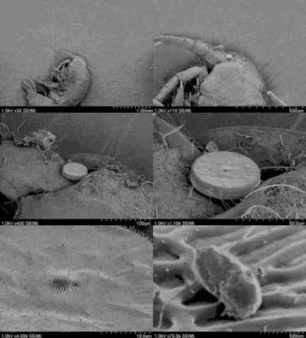
- A human hair attached to skin viewed from a microscope.

- Comparison of the tip of a hypodermic needle, vipers fang, spiders fang and the stinger of a scorpion

- Brooklynns board:-)

- Comparison Of The Tip Of A Hypodermic Needle, A Viper’s Fang, A Spider’s Fang And The Stinger Of A Scorpion.

- From left to right: The tip of a needle, a vipers fang, a spiders fang and the stinger of a scorpion.

- organic matter

- Macro Photography

- Instant Of Test Nuclear Detonation Captured By Harold Edgertons Rapatronic Camera With Shutter Speed Of One Hundred Millionth Of A Second. Circa 1950s. [1300 × 1051]

- Microscope pictures

- Microscopy

- Assembled

- Plant cell images

- Brain

- Decadence/Decay ...###

- Recycled Glass Countertops

- Body fluid

- Anatomy & Physiology/Medical Terminology

- A hibiscus cell through a microscope {OC}

- Clay supplies

- beverly high school

- The first few milliseconds after a nuclear bomb is detonated

- Anatomy And Physiology

- 31 Days Of Halloween

Soil is home to the greatest biodiversity found on earth. As Rattan Lal said - There is no life without soil and no soil without life!! Last year, we looked at soil and its benefits on a micro level. Pictured here, at 400x magnification, a network of mycorrhizal fungi (stained blue) is visible inside plant roots. In this symbiotic partnership, plants provide carbon-rich sugars in exchange for the fungus’s offering of nutrients and precious water scavenged from the soil. Soil is one of the key solutions to our climate crisis. Healthy soil traps carbon! Watch the 3 part series @patagonia how regenerative organic farming methods are changing the way we grow food and fiber and restore the health of our soil and mother earth. #regenerativeagriculture #regenag #organicfarming #organic #soilislife #soil #climatecrisis #carbon #farm #farming - @kerioberly on Instagram

- There are several cases where frogs, toads, and other small animals are found concealed within solid stone – alive. There are other instances too, where workers would cut down trees, and find hoards of frogs within the interior.

- Closeup of an old basketball

- disturbingly beautiful

- Biology 3

- books, articles, etc.

- Ballpoint Pen

- Marine diatoms, one of the most ecologically significant organisms. They are unicellular algae with transperant, opaline silica walls. Account for almost 20 percent of global photosynthetic fixation of carbon which is more than all of the Worlds tropical rainforests combined.

- Optical coherence tomography

- A sooted glass plate reveals the structure of the hydrogen/air detonation wave that has passed across it

- Forbidden grapes (staphylococcus bacteria)

- Saturn (Planet)

- Parakaryon myojinensis, a creature found only once near a hydrothermal vent south of japan, and is so unique that scientist cant agree if its a prokaryote (bacteria, etc), an eukaryote (animals, plants, fungi, etc) or a new domain entirely.

- 1-A-5 May

- BSO: Vakantie Sterrenjacht

- Hair science

- blood infection

- Smola

Keratoprosthesis is a surgical procedure where a diseased cornea is replaced with an artificial cornea. Traditionally, keratoprosthesis is recommended after a person has had a failure of one or more donor corneal transplants. More recently, a less invasive, non-penetrating artificial cornea has been developed which can be used in more routine cases of corneal blindness. While conventional cornea transplant uses donor tissue for transplant, an artificial cornea is used in the Keratoprosthesis procedure. The surgery is performed to restore vision in patients suffering from severely damaged cornea due to congenital birth defects, infections, injuries and burns. Keratoprotheses are made of clear plastic with excellent tissue tolerance and optical properties. They vary in design, size and even the implantation techniques may differ across different treatment centers. The procedure is done by ophthalmologists, often on an outpatient basis. Although many keratoprostheses have been developed, only four models are currently in commercial use: the Boston keratoprosthesis, Osteo-Odonto-Keratoprosthesis (OOKP), AlphaCor and the KeraKlear Artificial Cornea. Image: Mariagessa. #keratoprosthesis #cornea #eyedisease #eyesurgeon #ophthalmologist #ophthalmology #optometrystudent #optometrist #optometry #biomechanical #eyedoctor #eyemed #medicalschool #medicaldoctor #oculista #eyehealth #eyecare #orthoptist #orthokeratology - @optometry.case on Instagram

- Tissue biology

- First Look At The New Coronavirus Under A Microscope. Stay Safe Out There People

- Designer Chandeliers

- Sand viewed through a microscope is not what you would expect

- (OC) Photo of plant cells under the microscope.

- Best homeo

- soft ground. love.

- Astronomer here! I cross stitched a radio time lapse of Supernova 1987A from my PhD thesis! I made the figure for the paper itself, and all the scaling, coordinates, fluxes, etc is scientifically accurate in this stitch

- Gastrointestinal System

- Embryonic development

- The closest we can see DNA

- Coloring tutorial

- Abstract, Laser , STRING ART. DIFFERENT COLORS

- Embryonic development

- LA CIENCIA

- [ Dungeon ]

- bands

- Bigfoot sightings

- Dining Room Sofa

- uric acid

This is Erosion according to Matthew. White Statuary Marble Bass Relief 23.6x15.7x1.2in - 60x40x3cm. Rare Marble #micromegalicinscriptions DM for Acquisitions. - - - @matteomaurostudio on Instagram

- I herd u liek molecules. This one is napthalenetetracarboxylic diimide.

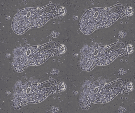
- That’s not a Cheeto. It’s a form of Protozoa.

- eyes

Cyanobacteria (blue-green algae) are found in a broad range of environments, from hot springs to marine and fresh waters, and soil crusts to polar icecaps. By understanding exactly how cyanobacteria harvest light and convert it to energy, scientists may be able to engineer highly efficient photosynthetic units to help increase crop yields. In a paper published recently in Nature Plants (doi.org/10.1038/s41477-020-0718-z) Vincenzo Mascoli (PhD student at Vrije Universiteit @vuamsterdam) and colleagues investigated cyanobacteria from shaded environments to learn more about the trade-offs between covering the largest portion of the solar spectrum for photosynthesis and achieving the highest yields of energy conversion. Vincenzo shares more about the work in a post for our Microbiology Community: go.nature.com/38UUTKK (link in bio). Image: Cells of a cyanobacterium, Chlorogloeopsis fritschii, under the microscope. Each cell is around 5 to 10 thousandths of a millimetre (5-10 μm) across. Credit: Luca Bersanini. #cyanobacteria #bluegreenalgae #biophysics #photosynthesis #microscopy #microbe - @nature.research on Instagram

- Microbiology

- Facial hair; razor vs electric shaver

- First photograph of a bacterium (anthrax), taken by Robert Koch in 1877

- Eclectic Oddities

- AAA

- Green algae

- BLUE

- Upper East Side Apartment

- alphabet images

- Lori Zimmerman, Selected Works

- I made a star trek logo out of atoms

- Whale sharks have dermal denticles (tiny teeth-like scales) on their eyes which protect them from abrasion. They can also pull their eyes back into their heads. It is hypothesized that these protective measures point to an importance in vision which has long been thought to not be the case in sharks

- Pus from husbands eye through the microscope looks like a cool planet.

Cortezas de Esperanza.. mis diseños #orfebreria #joyasplata #amoloquehago #quina #cascarilla 🦄💜🍀 @j.borrero_13 @d.dborrero @nicole_borrero - @s_o_f_i_a_adlt on Instagram

- Dulce et decorum est

- Looks like a fancy arts and craft project. But these are real snowflakes under an electron microscope

- Metalic texture

- Rain Gauge

- Art note !

Vanaf vandaag is de nieuwe museale opstelling ‘Corona gaat viral’ te bewonderen in Micropia. Met deze opstelling wil Micropia de huidige coronapandemie van achtergrondinformatie voorzien. Was het de vleermuis, het schubdier of de mens? Naast de bestaande opstellingen in Micropia met virusmodellen van hiv, ebola en het pokkenvirus is nu ook het coronavirus te zien. De nieuwe opstelling is een combinatie van een opgezet schubdier en een 3D-geprint natuurgetrouw model van het coronavirus. Dit model is gemaakt van kunststof. In werkelijkheid is het virus ongeveer 125 nanometer in doorsnede. Het virus is te herkennen aan de knuppelvormige stekels op het membraan waardoor het de naam ‘corona’ - Latijn voor ‘kroon’ - heeft gekregen. #corona #viral #micropia #amsterdam #virus #pandemie - @micropia_amsterdam on Instagram

- clay bowls

- Astronaut Costume

- Astronomy

- c. a

🇬🇧 When something happens... 💥 Shocking photo by our friend @and_sh 🔝 Thanks for the beautiful photo and Welcome to the group! 🙏 Do you want to participate with the community? Tag us with your cases and send us a DM! - - - #perioresto #beppro #periimplantitis #periimplantdiseases #surfacedecontamination #aestheticdentistry #adhesivedentistry #implantdentistry #dentalimplants #veneers #restorativedentistry #aesthetics #dentalart #dentalstudentproblems #teeth #implants #dental #dentalcases #bone #boneregeneration #odontologia #odontoiatria #implantologia - @perioresto on Instagram

- 1950s Art Moderne master bedroom

- dental

Abstract Painting Title: Untitled Medium: Acrylic on Canvas Board Size: 2.5 W x 2 .5 H (art dimension ) Frame Dimension: 6 W x 6 H . . #abstractpainting #abstractpainter #contemporaryart #dorociak #fineart #originalart #artistsofinstagram #smallart #painter #art #dorostudios - @dorostudios on Instagram

- My crystals of copper(II) hexafluoroacetylacetonate

- Cloth Nappies Tips

- HIV virus budding from the surface of a white blood cell

- Pet CT

- Audio-Visual

- Black and White Art

- ABCs

